
Technologies技术与方法
During the development of life sciences, microscopy has always played a vital role. With the emergence and improvement of new techniques, cutting-edge researches on structure and dynamic properties of complex biological systems such as biological macromolecular complexes at nanoscale, cells at micrometer scale, and even tissues and organs at larger scales are experiencing major breakthroughs, bringing clearer understandings on how life units complete advanced biological functions from cell division, neuronal connections, to human brain cognition and consciousness at multiple spatiotemporal scales.在生命科学的发展历程中,显微成像始终发挥着至关重要的作用。随着新技术的出现与完善,针对纳米尺度生物大分子复合物、微米尺度细胞以及更大尺度组织和器官等复杂生物系统的结构与动态特性的前沿研究正取得重大突破,使人们能够在多个时空尺度上更清晰地理解生命单元如何完成从细胞分裂、神经元联结到人脑认知与意识等高级生物功能。
By interdisciplinary collaborations, we focus specifically on effective technologies and methods for analyzing the structure and function of neural synapses and neuronal circuits, especially nanoscale imaging techniques such as CryoEM, Cryo-CLEM and super-resolution imaging. We also independently developed a new ultra-high-speed 3D fluorescence microscopy technology (VISoR), and combined it with electrophysiology and behavioral methods for high-throughput analysis of whole-brain structure and activity traces.通过跨学科合作,我们重点发展解析神经突触和神经环路结构与功能的有效技术和方法,尤其是冷冻电子显微、冷冻光电关联显微和超分辨成像等纳米尺度成像技术。我们还自主研发了超高速三维荧光显微成像技术(VISoR),并将其与电生理和行为学方法结合,用于全脑结构和活动痕迹的高通量分析。
Electron Microscopy电子显微学
Electron microscopy and correlative microscopy电子显微与关联显微
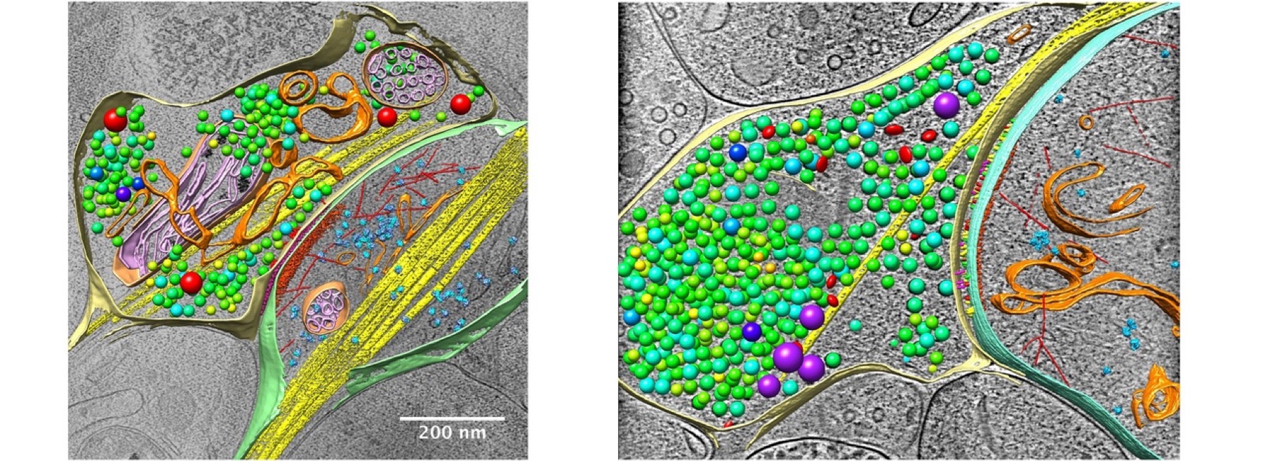
Electron microscopy, due to its extremely high resolution, has always been the main technique used for the analysis of tissue and cellular ultrastructure, molecular localization and distribution. In particular, the latest advances in cryo-EM, including cryo-EM tomography for 3D reconstruction and single-particle reconstruction, enabled us to resolve the ultrastructural details of subcellular and even protein macromolecular complex ultrastructure at nanometer and even atomic level resolution. At the same time, the evolving field of correlative microscopy combines optical and electron microscopy to achieve multi-scale correlated imaging of the same sample. By integrating the high resolution, high sensitivity, and high specificity, this technology enables us to observe and analyze the ultrafine structures such as protein complexes in subcellular systems including synapses with unprecedented vision, leading to a new understanding of various biological cell structures and functions.电子显微镜凭借极高的分辨率,一直是解析组织和细胞超微结构以及分子定位与分布的主要技术。特别是冷冻电镜领域近年来的进展,包括用于三维重构的冷冻电子断层成像和单颗粒重构,使我们能够在纳米乃至原子尺度解析亚细胞结构及蛋白质大分子复合物的超微结构。同时,持续发展的关联显微技术将光学显微与电子显微相结合,实现同一样品的多尺度关联成像。该技术综合高分辨率、高灵敏度和高特异性,使我们能够以前所未有的视角观察和分析突触等亚细胞系统中的蛋白质复合物等精细结构,从而深化对多种生物细胞结构与功能的认识。
Cryo-electron tomography, cryo-ET冷冻电子断层成像(cryo-ET)
We aim to resolve the 3D structures of various amorphous biological samples, including tissues, cells, subcellular components, and biomolecular complexes, under near-physiological conditions at nanometer resolution. The basic principle of CryoET involves rapidly freezing the sample to preserve its structures and placing it into CryoEM. By tilting the sample and collecting a series of 2D projection images, the 3D structure is reconstructed via the back-projection method. With the improvement of sample preparation technology and EM imaging quality, the quality of CryoET data has been greatly improved, enabling the in situ identification of macromolecular complexes. Combined with image processing techniques such as pattern recognition, the localization and distribution of protein molecules in specific samples, such as individual synapses, can be identified, so as to achieve quantitative analysis of the protein quantity and localization. Furthermore, by combining sub-tomogram averaging technology, it is possible to achieve high-resolution 3D structural analysis of biomolecules in situ with resolution approaching atomic level. With the development of technology, CryoET is becoming a primary research tool for the quantitative analysis of protein organization, localization and distribution at the cellular and subcellular levels, as well as in situ structural analysis of biological macromolecules.我们旨在近生理条件下,以纳米分辨率解析组织、细胞、亚细胞组分和生物分子复合物等多种非晶态生物样品的三维结构。cryo-ET的基本原理是快速冷冻样品以保存其结构,并将其置于冷冻电镜中;通过倾转样品采集一系列二维投影图像,再利用反投影方法重构三维结构。随着样品制备技术和电镜成像质量的提升,cryo-ET数据质量显著提高,可在原位识别大分子复合物。结合模式识别等图像处理技术,可以识别特定样品(如单个突触)中蛋白质分子的定位和分布,实现对蛋白质数量与定位的定量分析。进一步结合子断层平均技术,可在原位对生物分子进行接近原子分辨率的高分辨三维结构解析。随着技术发展,cryo-ET正成为在细胞与亚细胞层面定量分析蛋白质组织、定位和分布,以及原位解析生物大分子结构的重要研究工具。
Cryo-correlative light and electron microscopy, cryo-CLEM冷冻光电关联显微(cryo-CLEM)
Cryo-correlative light and electron microscopy (Cryo-CLEM) is materializing as a widespread approach amalgamating the advantages of both fluorescence light microscopy (FLM) as well as three dimensional (3D) cryo-electron tomography (cryo-ET) to reveal the ultrastructure of significant target molecules with specific cellular functions. Cryo-CLEM allows imaging of cells by means of fluorescence microscopy exhibiting the location of the destined molecule at high temporal and spatial resolution while cryo-ET is employed to analyze the 3D structure at a molecular resolution in close-to-physiological condition.冷冻光电关联显微(cryo-CLEM)结合荧光光学显微(FLM)与三维冷冻电子断层成像(cryo-ET)的优势,用于揭示具有特定细胞功能的重要靶分子的超微结构。cryo-CLEM先通过荧光显微成像,以较高的时间与空间分辨率确定目标分子的位置,再利用cryo-ET在接近生理的条件下以分子分辨率解析其三维结构。
Light Microscopy光学显微学
Light-Sheet Fluorescence Microscopy and VISoR光片荧光显微成像与VISoR
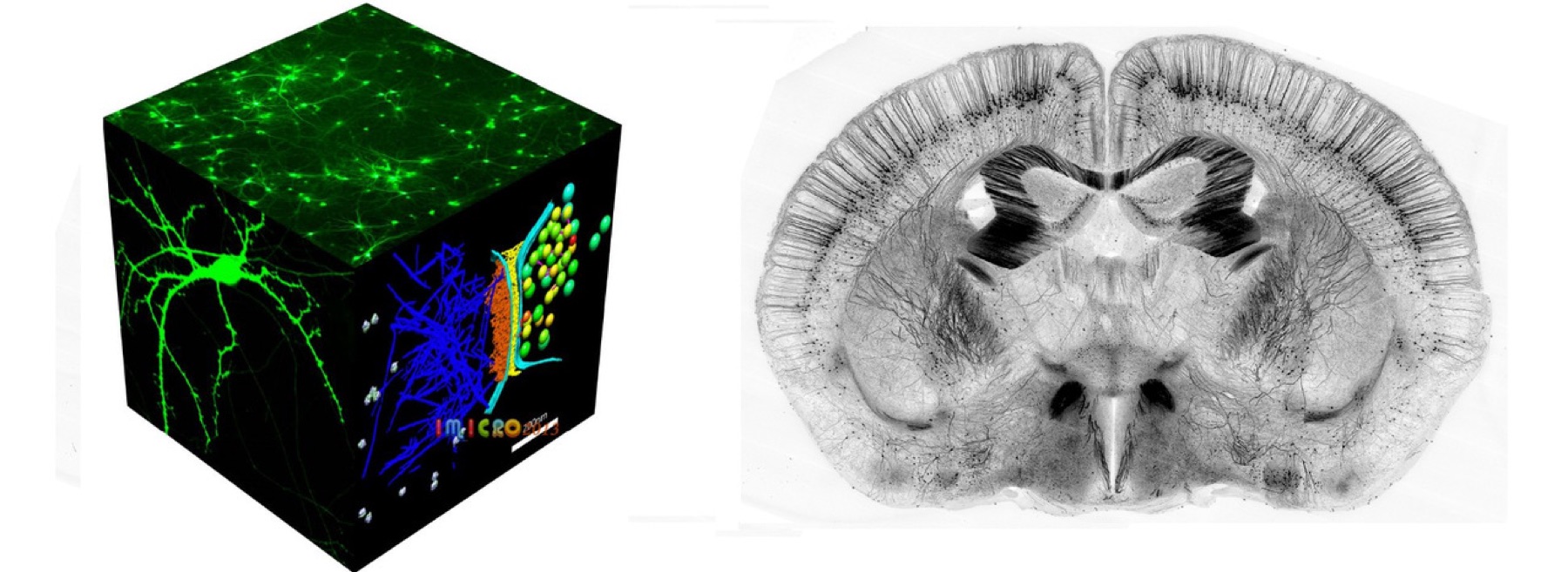
High-speed mesoscale imaging enables large-volume 3D visualization of tissues and entire organs at 1-5 μm resolution, supporting single-cell mapping and long-range projection tracing. Light-sheet fluorescence microscopy (LSM) serves as the primary modality for volumetric imaging of optically cleared transparent samples. A variety of LSM variants have been developed and are now broadly applied across diverse biological disciplines.高速介观成像能够以1至5微米分辨率对组织和完整器官进行大体积三维可视化,支持单细胞图谱绘制和长程投射追踪。光片荧光显微成像(LSM)是对光学透明化样品进行体积成像的主要方式。多种LSM变体已经发展起来,并广泛应用于不同的生物学领域。
LSM illuminates the specimen with a thin light sheet and collects the emitted fluorescence along a separate optical path, typically arranged orthogonally to the excitation plane. Because fluorescence excitation is confined to a thin tissue slice, the technique provides inherent optical sectioning. Moreover, this excitation-detection geometry offers high excitation energy efficiency, resulting in minimal photobleaching of bulk tissue. These characteristics make LSM an ideal modality for three-dimensional imaging, including whole-brain imaging of cleared tissues.LSM使用薄光片照明样品,并通过独立的光路采集发射荧光;激发光路与探测光路通常正交排列。由于荧光激发被限制在较薄的组织切面内,该技术天然具备光学切片能力。同时,这种激发与探测几何结构具有较高的激发能量利用效率,可将大体积组织的光漂白降至较低水平。这些特点使LSM成为三维成像的理想方式,包括对透明化组织进行全脑成像。
High-resolution imaging of large specimens generates massive datasets, with terabyte-scale datasets being typical and petabyte-scale becoming increasingly common. Throughput is therefore of paramount importance in such applications. To address this, we developed the VISoR system, a specialized light-sheet microscope, along with its associated sample processing workflow for high-speed volumetric imaging. In this system, both the excitation and detection arms are tilted above the specimen in a "V" configuration, which avoids any physical obstruction to the lateral translation of arbitrarily large specimens. The specimen is translated continuously during image acquisition, and a dedicated motion-correction technique eliminates motion blur caused by sample movement. This approach pushes sustained imaging throughput to the practical limit of the camera. Furthermore, the Blockface-VISoR method was developed for highly heterogeneous tissues, enabling subcellular-resolution imaging of whole mouse bodies. This method retains the high-throughput advantages of VISoR light-sheet microscopy while ensuring data integrity, and extends the capability of individual projection tracing from the whole brain to the entire body.大型样品的高分辨成像会产生海量数据,TB级数据已很常见,PB级数据也越来越多。因此,成像通量在这类应用中至关重要。为此,我们开发了专用光片显微镜VISoR及其配套样品处理流程,用于高速体积成像。该系统的激发臂和探测臂均倾斜置于样品上方,形成“V”形结构,避免对任意大小样品的横向移动造成物理阻挡。成像过程中样品连续移动,专用运动校正技术可消除样品移动引起的运动模糊,使持续成像通量达到相机的实际极限。此外,针对高度异质性组织,我们开发了Blockface-VISoR,可对小鼠全身进行亚细胞分辨率成像。该方法在保证数据完整性的同时保留VISoR光片显微成像的高通量优势,并将单神经元投射追踪能力从全脑扩展至全身。
VISoR microscopy is distinguished by its rapid volumetric imaging of cleared samples, and its hybrid design, Blockface-VISoR, further enables large-specimen and whole-body imaging. Collectively, these techniques constitute a versatile toolkit for mesoscale connectomics and single-neuron projection mapping in both rodents and primates, and have yielded the world's first high-resolution whole-brain dataset for monkey and whole-body dataset for mouse.VISoR显微成像的突出特点是能够对透明化样品进行快速体积成像,其混合设计Blockface-VISoR进一步支持大型样品和全身成像。这些技术共同构成适用于啮齿类和灵长类介观联接组学及单神经元投射图谱绘制的多用途工具体系,并已获得世界首个高分辨率猴全脑数据集和小鼠全身数据集。
Confocal laser scanning microscopy激光扫描共聚焦显微成像
In a conventional (i.e., wide-field) fluorescence microscope, the entire specimen is flooded evenly in light from a light source. All parts of the sample can be excited at the same time and the resulting fluorescence is detected by the microscope's photodetector or camera including a large unfocused background part. The principle of confocal imaging was patented in 1957 by Marvin Minsky and aims to overcome some limitations of traditional wide-field fluorescence microscopes. A confocal microscope uses point illumination (see Point Spread Function) and a pinhole in an optically conjugate plane in front of the detector to eliminate out-of-focus signal, the name confocal stems from this configuration.在传统的宽场荧光显微镜中,光源均匀照亮整个样品,样品各部分可同时受到激发,由显微镜的光电探测器或相机采集所产生的荧光,其中也包括大量失焦背景。Marvin Minsky于1957年为共聚焦成像原理申请专利,旨在克服传统宽场荧光显微镜的部分局限。共聚焦显微镜使用点照明,并在探测器前方的光学共轭平面放置针孔以消除失焦信号,“共聚焦”之名即源于这一结构。
Two-photon excitation microscopy双光子激发显微成像
Two-photon excitation microscopy (TPEF or 2PEF) is a fluorescence imaging technique that is particularly well-suited to image scattering living tissue of up to about one millimeter in thickness. Unlike traditional fluorescence microscopy, where the excitation wavelength is shorter than the emission wavelength, two-photon excitation requires simultaneous excitation by two photons with longer wavelength than the emitted light. The laser is focused onto a specific location in the tissue and scanned across the sample to sequentially produce the image. Due to the non-linearity of two-photon excitation, mainly fluorophores in the micrometer-sized focus of the laser beam are excited, which results in the spatial resolution of the image. This contrasts with confocal microscopy, where the spatial resolution is produced by the interaction of excitation focus and the confined detection with a pinhole. Due to the multiphoton absorption, the background signal is strongly suppressed, leading to an increased penetration depth for this technique. Two-photon excitation can be a superior alternative to confocal microscopy due to its deeper tissue penetration, efficient light detection, and reduced photobleaching.双光子激发显微成像(TPEF或2PEF)是一种荧光成像技术,特别适合对厚度约一毫米以内的散射活体组织进行成像。不同于激发波长短于发射波长的传统荧光显微成像,双光子激发需要两个波长长于发射光的光子同时激发。激光聚焦于组织中的特定位置并在样品上扫描,依次生成图像。由于双光子激发具有非线性,主要只有位于微米尺度激光焦点内的荧光团被激发,从而形成图像的空间分辨率。这与通过激发焦点和针孔限制探测共同形成空间分辨率的共聚焦显微成像不同。多光子吸收能够显著抑制背景信号,因此该技术具有更大的穿透深度。凭借更深的组织穿透、较高的光收集效率和较少的光漂白,双光子激发在一些应用中优于共聚焦显微成像。
Super-resolution fluorescence microscopy超分辨荧光显微成像
Super-resolution microscopy is a series of techniques in optical microscopy that allow such images to have resolutions higher than those imposed by the diffraction limit, which is due to the diffraction of light. There are two major groups of methods for super-resolution microscopy in the far-field that can improve the resolution by a much larger factor: Deterministic super-resolution: the most commonly used emitters in biological microscopy, fluorophores, show a nonlinear response to excitation, which can be exploited to enhance resolution. Such methods include STED, GSD, RESOLFT and SSIM. Stochastic super-resolution: the chemical complexity of many molecular light sources gives them a complex temporal behavior, which can be used to make several nearby fluorophores emit light at separate times and thereby become resolvable in time. These methods include Super-resolution optical fluctuation imaging (SOFI) and all single-molecule localization methods (SMLM), such as SPDM, SPDMphymod, PALM, FPALM, STORM, and dSTORM.超分辨显微成像是一系列使光学显微图像分辨率突破光衍射极限的技术。远场超分辨显微成像主要有两大类方法,可显著提升分辨率。确定性超分辨方法利用生物显微成像中常用荧光团对激发光的非线性响应来增强分辨率,包括STED、GSD、RESOLFT和SSIM等。随机性超分辨方法则利用多种分子光源复杂的时间行为,使相邻荧光团在不同时间发光,从而在时间上被分辨。这类方法包括超分辨光学涨落成像(SOFI)以及SPDM、SPDMphymod、PALM、FPALM、STORM和dSTORM等单分子定位显微方法(SMLM)。
Stochastic optical reconstruction microscopy (STORM), photo activated localization microscopy (PALM), and fluorescence photo-activation localization microscopy (FPALM) are super-resolution imaging techniques that utilize sequential activation and time-resolved localization of photoswitchable fluorophores to create high resolution images. During imaging, only an optically resolvable subset of fluorophores is activated to a fluorescent state at any given moment, such that the position of each fluorophore can be determined with high precision by finding the centroid positions of the single-molecule images of a particular fluorophore. One subset of fluorophores is subsequently deactivated, and another subset is activated and imaged. Iteration of this process allows numerous fluorophores to be localized and a super-resolution image to be constructed from the image data.随机光学重构显微成像(STORM)、光激活定位显微成像(PALM)和荧光光激活定位显微成像(FPALM)利用光开关荧光团的顺序激活和时间分辨定位来生成高分辨率图像。成像时,每一时刻仅激活一组在光学上可分辨的荧光团,通过确定单分子图像的质心位置,高精度定位各荧光团。随后关闭这一组荧光团,再激活并成像另一组。重复这一过程即可定位大量荧光团,并由图像数据重构超分辨图像。
STORM has also been extended to three-dimensional imaging using optical astigmatism, in which the elliptical shape of the point spread function encodes the x, y, and z positions for samples up to several micrometers thick, and has been demonstrated in living cells. To date, the spatial resolution achieved by this technique is ~20 nm in the lateral dimensions and ~50 nm in the axial dimension.STORM还可通过光学像散扩展到三维成像,其中点扩散函数的椭圆形状编码数微米厚样品中的x、y、z位置。该技术目前实现的空间分辨率约为横向20纳米、轴向50纳米。
Behavioral Studies行为学研究
Behavioral Paradigms行为学范式
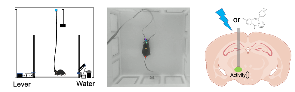
We mainly use operant learning paradigms, Go/No-Go learning paradigms, and cooperative learning paradigms to study the neural circuit mechanisms underlying learning and memory in mice. Our long-term goal is to uncover general principles of learning and memory that operate across brain regions and behavioral tasks.我们主要采用操作性学习范式、Go/No-Go学习范式、合作学习范式等,研究小鼠在不同学习记忆场景下的神经环路机制,最终希望揭示大脑跨脑区、跨任务的通用学习记忆原理。
Behavioral Analysis行为分析
We use tools such as Bonsai, PsychoPy, DeepLabCut, and VisuTrack to acquire and analyze mouse behavioral information. These approaches allow us to perform detailed behavioral classification and kinematic analysis, capturing fine-scale dynamic changes during learning.我们利用Bonsai、PsychoPy、DeepLabCut和VisuTrack等工具,采集和分析小鼠行为信息,进行深入的行为分类和动力学分析,捕捉学习过程中的精细动态变化。
Optogenetics光遗传学
Optogenetics uses engineered light-sensitive proteins, or opsins, to control neuronal activity with light. By expressing these proteins in specific neuronal populations, we can precisely regulate ion flux across the neuronal membrane and thereby promote depolarization or hyperpolarization, leading to activation or inhibition of selected brain regions or neural circuits. We use this approach to test the causal roles of candidate brain regions and neural circuits in mouse learning and memory.光遗传学利用光敏蛋白调控神经元活动。通过遗传工程手段将光敏蛋白表达在特定神经元群体中,并用光精确控制细胞膜上的离子通道或离子泵,从而引发神经元去极化或超极化,实现对特定脑区或神经环路的激活或抑制。利用该方法,我们可以研究在小鼠学习记忆过程中可能起关键作用的脑区或神经环路的功能机制。
Chemogenetics化学遗传学
Chemogenetics uses engineered G protein-coupled receptors that can be activated or inhibited by specific ligands, such as CNO or related compounds. By expressing these receptors in defined neuronal populations, we can pharmacologically modulate the activity of specific brain regions or neural circuits and examine the resulting effects on neural activity and behavior. We use this method to investigate the functional mechanisms of brain regions and neural circuits that may play key roles in mouse learning and memory.化学遗传学通过改造G蛋白偶联受体,使其能够被特定配体,如CNO或相关化合物,激活或抑制。将这些受体通过遗传工程手段表达在特定神经元群体中后,可以通过药物调控特定脑区或神经环路的活动,并观察其对神经活动和行为表型的影响。利用该方法,我们可以研究在小鼠学习记忆过程中可能起关键作用的脑区或神经环路的功能机制。
Neural Activity Recording神经活动记录
Neural Activity Recording Technologies神经活动记录
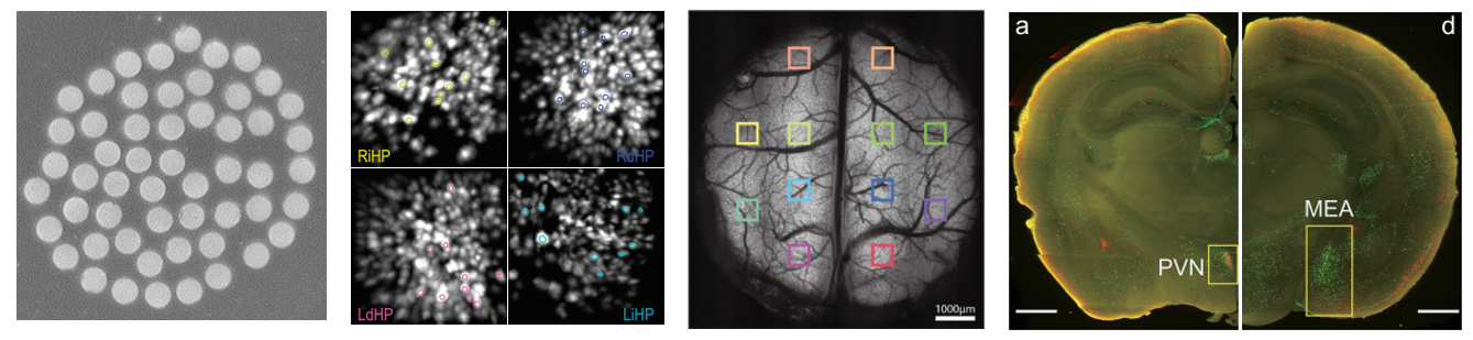
We use a range of neural activity recording and manipulation approaches to investigate neural activity patterns during mouse learning, with a particular focus on the transition period before and after learning occurs.我们利用多种神经活动记录与调控技术,研究小鼠学习过程中的神经活动模式,尤其关注小鼠刚学会任务前后的神经活动变化。
Multichannel Fiber Photometry System多通道光纤记录系统
Using genetically encoded neural activity indicators, such as GCaMP, we target multiple optical fibers to different brain regions. Signals from these fibers are acquired synchronously with a camera-based system, allowing us to monitor time-resolved population neural activity across multiple brain areas in freely moving mice. We use this system to investigate dynamic multi-region neural activity patterns across different stages of learning.基于基因编码的神经活动探针(如GCaMP),将多根光纤靶向不同脑区,利用相机同步采集多根光纤中的信号变化,进而获取自由运动状态下动物大脑内不同区域的实时神经元群体活动信息。我们利用该系统研究小鼠在学习的不同阶段的多脑区动态神经活动模式。
Ultracompact Head-Mounted Microscopy System超紧凑头戴式显微成像系统
We have developed an ultracompact head-mounted microscopy system that integrates miniature optoelectronic components and CMOS image sensors into a device weighing less than 0.5 g. This system enables neural activity imaging from up to four brain regions in freely moving mice. With this platform, we can study neural activity patterns within and across brain regions during learning.我们自主开发了超紧凑头戴式显微成像系统,将微型光电元件、CMOS图像传感器等集成在重量不到0.5克的头戴式设备中,在小鼠中实现了自由活动状态下最多4脑区神经元活动成像。利用此系统,我们可以研究小鼠在学习过程中不同脑区内的神经元活动模式。
Large-Field Two-Photon Imaging System大视场双光子成像系统
Two-photon imaging offers low background, high spatial resolution, and optical sectioning capability. Our system provides a field of view up to 8 mm in diameter, covering large areas of the mouse dorsal cortex. It enables high-quality recording of neural activity across broad cortical regions in head-fixed mice, as well as high-resolution imaging of local neuronal and synaptic dynamics under appropriate imaging configurations. We use this system to study neuronal and synaptic activity patterns across cortical areas during specific behavioral tasks.双光子成像具有背景低、空间分辨率高和光学切片能力强等优势。该系统具有最大直径8 mm的视野范围,可覆盖小鼠背侧皮层的大范围区域,能够在头部固定状态下获取小鼠大脑大范围、跨皮层区域的高质量神经元活动信息;在适当的高分辨率成像配置下,也可用于记录局部神经元和突触动态变化。利用该系统,我们可以研究小鼠在特定任务范式下不同皮层区域的神经元或突触活动模式。
Whole-Brain Neural Activity Mapping全脑神经活动印迹图谱
Using our self-developed rapid three-dimensional imaging technology (VISoR) and whole-brain immunofluorescence staining methods, we can label and map activity-associated neuronal states across the entire brain within defined time windows, such as approximately 1.5 ± 0.5 hours before brain collection. For example, immediate early genes such as c-Fos can be used as markers of recent neuronal activity. This approach allows us to compare whole-brain neural activity patterns across different learning stages, such as before and after task acquisition.利用自主研发的快速三维成像技术VISoR和全脑免疫荧光染色方法,我们可以在特定时间窗口内,例如取出大脑前约1.5 ± 0.5小时,对全脑范围内的活动相关神经元状态进行标记和成像。例如,可利用即刻早期基因c-Fos作为近期神经元活动的标记,获取该时间窗口内的全脑神经活动相关图谱。利用该技术,我们可以比较小鼠在不同学习阶段,如学会前和学会后的全脑神经活动状态。